Investigations
Ultrasound
An ultrasound (or Duplex) is a scanning method that uses high frequency sound waves to assess patients with vascular disease. It shows blood vessels (arteries and veins) in real time on a computer screen. It can also demonstrate blood flow through the vessels in colour (Doppler) which can be accompanied by sound. Ultrasound is non-invasive so there are no painful tubes or needles that need to be inserted into the body. It doesn’t require the use of potentially harmful dye or radiation and there are no known side-effects.
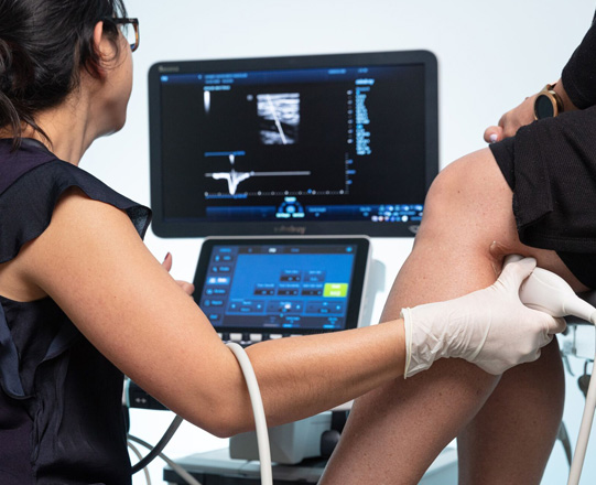
An ultrasound is an operator-dependent examination so that its reliability depends on the experience and knowledge of the sonographer performing the scan and the Specialist interpreting the results. With modern advanced technology providing high quality images, a reliable sonographer can use their valuable years of experience and knowledge to identify vascular problems. Ultrasound changes can be subtle and potentially missed with older equipment and inexperience.
An ultrasound can identify the location and severity of narrowed arteries (atherosclerosis), aneurysms (dilated arteries), blood clots in veins (thrombosis) and abnormal blood flow through diseased varicose veins. As well as identifying these conditions, it can also be used to monitor for disease progression in patients not yet requiring treatment (surveillance), and to follow-up patients who have undergone a vascular procedure to ensure that the treatment remains effective.
An ultrasound is the best method to initially investigate patients with suspected vascular disease however some patients may require more detailed studies such as angiography or computerised tomography (CT) scanning in order to plan any required treatment. These other tests are however are more invasive and carry some risk and ideally should not be arranged before seeing a vascular surgeon. Dr Freeman can provide advice in relation to whether there is a need for these additional tests to be carried out.
Little or no special preparation is usually required for an ultrasound unless the ultrasound is around the abdomen (tummy) as gas in the bowel can interfere with ultrasound transmission. For this reason patients should ideally have nothing to eat (fast) after midnight for abdominal scans which are normally performed in the morning. You can however have unlimited water and take all your medications.
If having a test for varicose veins do not wear compression stockings for 24 hours. Do not apply moisturisers on the day of the examination. Any bandages used for ulcers will need to be removed before attending your appointment and a clean simple dressing applied. If possible leave or remove all jewellery at home and wear loose, comfortable clothing to enable the sonographer to gain access to the targeted area for ultrasound.
Angiography
An angiogram is an x-ray procedure that involves the injection of a dye (contrast) into blood vessels usually through the groin at the top of your thigh. It provides very high-quality X-ray images of blood vessels including information regarding blood flow through the vessels. It does have some risks. These include bruising, bleeding or injury to the artery that is accessed. Ultrasound is used at the time of accessing the vessel to help avoid these complications. It is also important that someone experienced places pressure on the access site in the groin and patients spend time in bed after the procedure to help prevent complications such as bleeding from the puncture site.
Some patients have minor reactions to the dye such as a rash or itching and some are allergic to the dye used for angiography, however severe reactions are rare. Newer dyes are safer than those used in the past, people at risk of having an allergic reaction can be given steroids to minimise any allergic reactions. In patients with a history of kidney problems, diabetes or heart disease, the dye can cause further kidney damage so as a precaution intravenous fluids for hydration are used to help prevent this. Some medications may need to be stopped.
As an alternative to dye, carbon dioxide can be used to prevent allergic reactions and kidney damage in at risk patients however the use of carbon dioxide can cause some discomfort and the images are not as good. Angiography does require exposure to radiation.
Angiography is used during minimally invasive (endovascular) procedures on blood vessels. It allows the observation and manipulation of devices used to treat the blood vessels in real time as the devices are passed through the vessel.
Procedures are usually done under local anaesthetic and patients will need to stay still in order to ensure good images are obtained. After these procedures the access vessel in the groin may need to be repaired with a special device. The details of this device are usually provided to the patient. In rare cases the device may become infected requiring it to be removed and the artery directly repaired
Patient’s need their kidney function checked with a blood test and need to confirm with the hospital any fasting (stop eating) requirements. The procedure and recovery period takes approximately six hours in total. Arrangements should be made for someone to pick you up after the procedure, do not drive a vehicle or take public transport after the procedure. Please ensure you have someone to stay with you for at least two hours after returning home in case of any unexpected bleeding from the puncture site.
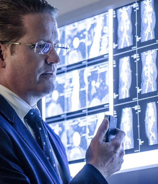
Computed tomography scanning is a series of x-ray images taken by a machine as it rotates around a patient. These x-rays are fed into software, which then can create an image of the vascular system. This can identify diseased arteries and veins, determine the severity of the problem and assist with planning treatment. Patient’s having a CT scan to examine their veins and arteries will usually need an IV injection of a dye (contrast) into a vein in your arm so there is the risk of allergic reaction or kidney damage. It does also require exposure to radiation. It doesn’t provide information about blood flow and there can be problems if the vessels are very diseased or small as is the case below the knee.
Specialists, sonographers and radiographers performing angiography and computed tomography should have a clear understanding of the risks involved with radiation exposure to the patient. There are several things they can do to reduce the radiation dose and accordingly the risks associated with radiation. You should discuss this with Dr Freeman.