Minimally Invasive Procedures
Dr Freeman has extensive experience in performing minimally invasive vascular procedures in appropriately selected patients. Minimally invasive procedures allows your surgeon to use techniques that limit the size and number of incisions (cuts), that they need to make to access arteries and veins. It’s typically considered safer than open surgery and you’ll usually recover more quickly, spend less time in the hospital, and feel more comfortable while you heal.
While these procedures potentially carry less risk than open vascular surgery, all procedures carry some risk. The information provided here is for general educational purposes only. Please discuss with Dr Freeman whether a minimally invasive vascular procedure is appropriate to treat your individual condition.
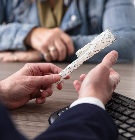
These days most aneurysms can be repaired using keyhole surgery through the groins. This is called endovascular aneurysm repair (EVAR). Stents are used to reline the aorta and protect the aortic wall from rupture. Endovascular aneurysm repair is effective and safe in selected patients with fewer risks than open surgery. It is still however a major operation performed under general anaesthesia in hospital. Complications may include injury to the kidneys, groin arteries used to access the aneurysm, infection or a leak can develop around the stents used to treat the aneurysm. In some cases patients may require additional procedures.
The development of minimally invasive keyhole surgery (endovascular surgery) through the groin restores blood flow through angioplasty, where balloons are used to open the arteries, and stenting, where a scaffold is used to hold arteries open. A patients’ suitability for endovascular surgery depends on a detailed examination of the arteries using angiography where dye is injected into the arteries. Complications can occur including damage to the groin arteries accessed during the procedure and kidney damage from the dye. The procedure requires exposure to radiation.
As an alternative to carotid endarterectomy (surgery to clean out the artery in the neck), stenting is a newer procedure that involves placing a scaffold across the narrowing in the carotid artery in the neck in order to prevent stroke. It is carried out under local anaesthetic through the groin. In addition to stroke, complications of stenting include damage to the artery in the groin used to place the stent. Not all patients are suitable for stenting. In addition to ultrasound further testing is usually required to determine if stenting is a reasonable alternative.
Patients with blocked mesenteric arteries supplying blood to the bowel and blocked renal arteries supplying blood to the kidneys may require the blood flow be restored. This can be achieved with stenting where a scaffold is inserted under angiography guidance to open the artery and clear the blockage. Not all patients with mesenteric or renovascular disease require a stent. After stenting patients need to be on aspirin (a blood thinning medication) and smokers should quit or seek help from your GP.
Arteriovenous fistulas used for dialysis can have problems that may require further treatment or surgery. They can develop narrowing that may limit blood flow and fistulas may block. Ultrasound surveillance is used to identify complications early so that they can be addressed, and the fistula flow maintained. This is done under local anaesthetic using angioplasty, where balloons are used to open the fistula, and stenting, where a scaffold is used to hold fistula open.
In patients with hyperhidrosis (excessive sweating), an operation to directly cut the nerves in the chest responsible for activation of the sweat glands in the hand and arm pits (thoracoscopic sympathectomy) is a permanent solution for patients. With any procedure comes risks, these can include excess sweating elsewhere (compensatory hyperhidrosis), air in the chest (pneumothorax), bleeding and inadvertent damage to other nerves.
As an alternative to surgical stripping of the veins, most patients with varicose veins or venous ulcers can be managed these days with endovenous ablation. A catheter is placed into the diseased vein under ultrasound guidance and activated to shut the vein down. The vein is effectively destroyed and as it heals the vein disappears. This has the advantage over surgical stripping of being less invasive. A cut in the groin is avoided, the procedure is less painful than surgery and patients return to normal activity after a shorter period.
A more recent treatment for varicose veins that has become available is glue (cyanoacrylate). This can be performed in clinic rooms under local anaesthetic. Like catheter and injection treatment it is minimally invasive and patients can return to normal activity almost immediately, however not all patients are suitable for glue. This is largely dependent on a patient’s ultrasound findings as to whether this is an appropriate treatment. Like catheter ablation some patients may require supplementary sclerotherapy.
Injection sclerotherapy involves injecting the vein with a sclerosant (an irritant solution) in order to shut the vein down. The sclerotherapy solution causes the vein to scar, forcing blood to reroute through healthier veins. The collapsed vein is reabsorbed into local tissue and eventually fades. Injection sclerotherapy is performed in the clinic rooms and is sometimes performed under ultrasound. Patients will need to wear a compression stocking on their leg for a period after the procedure but can walk, drive and essentially return to normal activity immediately following the procedure. Patients may experience soreness and lumps at the injection sites which usually settles over time and some patients develop staining which may result from trapped blood following injections. Multiple treatments may be required to achieve the results you are expecting.
Some patients with deep vein thrombosis develop an especially severe swollen leg. The patients that develop this may be appropriate for a procedure where a catheter is inserted to deliver direct clot-dissolving medication to the deep vein thrombosis (thrombolysis). A mechanical device can also assist in directly removing the clot from the deep veins. These procedures do carry some risks, particularly bleeding, and not all patients are suitable for this treatment, Dr Freeman can advise you on the best treatment options.
Some patients require insertion of a filter between a deep vein thrombosis and the lungs in order to stop clot travelling to the lungs. This filter is placed in the vena cava, the main vein in the abdomen. This is especially the case in patients who cannot have blood thinning medication or in whom the blood thinning medication is ineffective. Patients suitable for a vena cava filter can have the filter removed once it is no longer required.
In patients with pelvic congestion syndrome ovarian vein reflux can be responsible for pelvic pain and heaviness. To reduce the pressure in the pelvis ovarian vein coil embolization is used to stop the blood refluxing back down into the pelvis enabling patients to get relief from their pain. Immediately after the procedure the pain may feel worse, this normally settles over a period of days. The procedure is done under local anaesthetic through the groin and patients go home on the same day.
Open Vascular Surgery
Dr Freeman has extensive experience in performing open vascular surgery in appropriately selected patients. All surgical operations carry some risk however the information provided here is for general educational purposes only. Dr Freeman will discuss on an individual case by case basis whether open vascular surgery is appropriate to treat your specific condition and the risks involved.
Some patients benefit from surgery to clean out the carotid artery in the neck (carotid endarterectomy) in order to prevent stroke. The decision perform surgery is based on several factors including the degree of narrowing and whether the patient has had symptoms. Surgery on the carotid arteries does carry with it the risk of causing a stroke. This needs to be balanced against the risk of stroke without surgery which is particularly high in patients who have already suffered damage to the brain. Surgery also carries with it the risk of injury to the nerves that control the tongue, speech and swallowing. These nerves usually recover. There is also the risk of bleeding, especially given that patients need to continue aspirin around the time of surgery. Infection rarely occurs and in some patient’s blood pressure is difficult to control after surgery.
In the past all aneurysms were repaired by open surgery through a large incision in the abdomen. This involves directly sewing a graft into the patient to replace the aneurysm. This carries with it risks. As with any surgery infection and blood clots can rarely occur. Patients receive medication to help prevent these complications. Stress is placed on the heart so heart attack can occur particularly in patients with heart problems. Damage can be done to the kidneys. Very rarely someone might not survive the procedure due to complications.
Surgery for peripheral arterial disease has traditionally involved an open vascular surgical bypass to restore flow beyond blocked arteries in the legs. A bypass is a major procedure. Careful consideration needs to be given in relation to the risks versus the benefits. There can be problems with infection, the wounds, developing blood clots, the heart and lungs and bypasses can themselves block especially in patients who continue to smoke.
Patients requiring long term haemodialysis are best served with an arteriovenous fistula. These are surgically created by joining an artery to a vein, usually at the wrist or the elbow. After the operation and over a period of 6 to 12 weeks the blood flow through the fistula increases to the point where there is enough flow to support haemodialysis. Once this is the case the fistula can be accessed with needles through the skin and into the vein. Blood is then taken to and from an artificial kidney.
Thoracic outlet syndrome occurs when the nerves or vessels (veins and arteries) to the arm are compressed as they come out of the chest, go over the first rib and pass under the clavicle bone. Some patients are suitable for surgery to create more room for these structures. This can involve removing extra (cervical) ribs or alternatively the first rib. Damaged arteries and veins may also require surgical repair
While open surgery has largely been replaced by minimally invasive procedures, it remains appropriate for selected patients with varicose veins. Surgery involves directly removing diseased veins from the thigh and leg through incisions. Often an incision is placed in the groin where the vein is tied off before being stripped from the leg. Patients can have bleeding, pain related to the incisions and there is a risk of wound infection. Patients normally require a period off work and need to avoid any heavy or strenuous activity.